CORNEA
The cornea clings tightly to the front part of the eyeball like a transparent window. It is largely responsible for light refraction and thus the focusing of light, ensuring that a sharp image is created within the eye. Opacities in the cornea will usually adversely affect the person’s visual acuity in some way. Such opacities can be congenital or inherited, or arise as a result of inflammation or injury to the eye, and their effects can extend throughout the cornea or be restricted to individual layers (upper, middle or lower layers). Such changes to the cornea can be resolved via surgery, to ensure that an improved quality of visual acuity can be restored.
In the case of keratoconus, a central, conic deformation of the cornea occurs, causing it to become increasingly thin and unstable. This disorder often develops in phases. As it proceeds, the distortion of the cornea is often so strong and irregular that it cannot be compensated for by glasses or a contact lens and visual acuity is several reduced.
CORNEAL TRANSPLANTATION
While treatment formerly involved the cutting out of a central, circular area through the entire cornea and the suturing in of a disk of donor cornea (PKP, penetrating keratoplasty), the latest and most modern techniques enable the replacement (lamellar keratoplasty) or transplantation (DALK, deep anterior lamellar keratoplasty; DSAEK, Descemet’s stripping automated endothelial keratoplatsy; DMEK, Descemet’s membrane endothelial keratoplasty) of only those layers of the cornea actually affected. As a specialised partner cooperating with a number of health insurers, we perform this operation as day surgery without a hospital stay.
UV CROSS-LINKING
Corneal collagen cross-linking is used to treat an existing case of keratoconus. UV cross-linking treatments involve the drop-wise application of riboflavin to the patient’s cornea followed by UVA illumination. This stimulates cross-linking of the collagen fibres in the cornea, thus achieving a stabilisation or stiffening of the corneal tissue. In early-stage keratoconus, the technique can postpone progression for several years or cause it to cease completely.For UV cross-linking treatments, we use the new UV-X 2000 Crosslinking System, which utilises an innovative illumination profile adjusted to the corneal deformation to produce a more uniform cross-linking of the collagen fibres. Treatment time is 10 minutes and has thus been reduced to one-third of the illumination time previously required, guaranteeing maximum patient comfort.
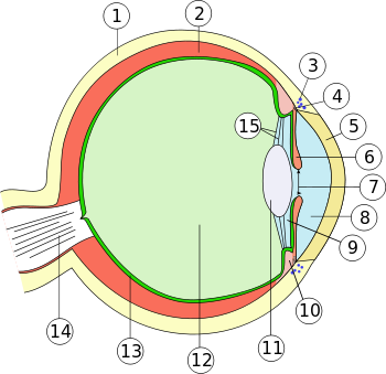
Cross-section through the human eyeball:
1. Sclera
2. Choroid coat
3. Canal of Schlemm (scleral venous sinus/scleral venous plexus)
4. Iris root (radix iridis)
5. Cornea
6. Iris
7. Pupil
8. Anterior segment (camera anterior bulbi)
9. Posterior segment (camera posterior bulbi)
10. Ciliary body (corpus ciliare)
11. Lens
12. Vitreous (corpus vitreum)
13. Retina
14. Optic nerve (nervus opticus)
15. Zonular fibres (fibrae zonulares)
Outer coat (tunica externa bulbi): 1. + 5.
Middle coat (tunica media bulbi, uvea): 2. + 6. + 10.
Inner coat (tunica interna bulbi): 13